Clear MRI but Neurological Symptoms? Why You Still Feel Sick
- Published
- No Comments
- Updated September 29, 2026
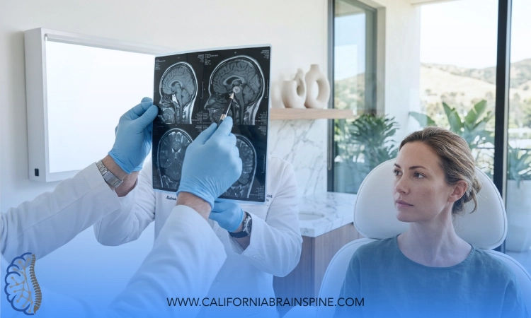
A normal MRI answers a question about structure. It says nothing about how the brain is working. Here is why a clear MRI but neurological symptoms is such a common pairing, what the scan can and cannot see, and what a functional evaluation adds.
The sentence people bring me is always some version of the same one. "The MRI was normal, so they said there is nothing wrong." And yet the person saying it is dizzy in the supermarket, foggy by two in the afternoon, sensitive to the light in their own kitchen, and not the person they were a year ago. A clear MRI but neurological symptoms is what a structural test looks like when the problem is one of function, and there is no contradiction in it.
I am Dr. Alireza Chizari, DC, DACNB, a board-certified chiropractic neurologist, and functional neurology at California Brain & Spine Center in Calabasas is built around exactly this gap. The scan has done its job and found no bleed, no tumour, no lesion. Nobody has yet measured how the eyes track, how the inner ear answers movement, how the heart rate behaves on standing, or how the balance system holds up with the eyes closed. Those are the measurements that explain most of the people who arrive with a normal report and a bad year behind them.
Key Takeaway
An MRI shows soft tissue and structure in detail; it does not measure how the nervous system performs. A concussion's acute symptoms are due primarily to a functional disturbance rather than structural injury, and persistent postural-perceptual dizziness shows frequently normal imaging. A clear scan rules out what it can see. A functional neurology evaluation measures what it cannot.
Can You Have a Clear MRI but Neurological Symptoms?
Yes. MRI shows soft tissue clearly and can help tell healthy from unhealthy tissue, which makes it the right test for structural problems. A concussion's acute symptoms are due primarily to a functional disturbance rather than structural injury, and persistent postural-perceptual dizziness shows frequently normal imaging. In conditions like those, a clear MRI but neurological symptoms is the expected pairing.
What an MRI shows, and what it does not
MedlinePlus, the National Library of Medicine's patient resource, describes an MRI as an imaging test that uses magnets, radio waves, and a computer to create detailed pictures of the inside of your body, one that shows organs and soft tissues clearly and can help tell healthy from unhealthy tissue in many cases. After a head injury it is a genuinely powerful tool. The National Institute of Neurological Disorders and Stroke notes that CT can show a skull fracture, bruising, bleeding or swelling, and that MRI is more sensitive and can pick up more subtle brain changes that a CT scan may miss. When something is structurally wrong, an MRI can help find it in many cases.
What it produces is a picture. A picture can show that a structure is present, intact and the expected shape. It cannot show whether the circuits running through that structure are timing correctly, whether the eyes and the inner ear are agreeing, whether the autonomic system is holding blood pressure on standing, or whether the balance system is leaning too hard on vision. Those are performances, and a performance does not photograph.
Why "normal" is a statement about structure
The clearest way I know to say this comes from the NCBI Bookshelf's review of concussion: a concussion is a traumatically induced transient disturbance of brain function, and its acute symptoms are due primarily to a functional disturbance rather than structural injury. Read that beside a normal scan and the apparent contradiction dissolves. The MRI was asked whether the structure was damaged, and it answered honestly. The symptoms come from how the structure is working, which was never the question the scan was built to answer. That is the functional gap, and it is where the rest of this article lives.
One thing has to be said before going further, because the search that brought you here is crowded with a different worry. Whether a disease could still be present despite a clear report, whether the scan should be repeated or read again, and whether a diagnosis such as multiple sclerosis applies are medical neurology's questions, and a functional evaluation is not a substitute for asking them. What a functional evaluation does is measure the performance that a clear scan leaves unmeasured, alongside medical care rather than instead of it.
Is It Normal to Feel Weird After an MRI?
For the scan day itself, MedlinePlus describes an ordinary experience: lying very still inside a loud, tunnel-like machine, sometimes with contrast through a vein, and after most MRI scans you can return to normal activities right away. Feeling weird after an MRI that outlasts the day, or that was there before the scan, is a different question.
What the scan day itself can do
People search this at two in the morning after a scan, and the honest answer has two halves. MedlinePlus describes the experience plainly: you lie on a table that slides into a large, tunnel-like machine, the machine makes loud noises as it takes pictures, the scan can take some time depending on the part of the body examined, you have to stay very still, and sometimes contrast is given through a vein beforehand. For most people that is unpleasant and then over: after most MRI scans you can return to normal activities right away, and if you were given medicine to help you relax, someone needs to take you home. Anything specific to contrast, to the medicine, or to a reaction on the day belongs with the imaging centre and the doctor who ordered the scan, not with this article.
When the feeling was there before the scan
The other half is the one I see. The person did not feel weird because of the MRI. They felt weird for months, the MRI was ordered to find out why, and it came back normal, which is why they are now typing "feeling weird after MRI" into a search box at night. If that is you, the scan has not caused anything and has not missed the point either. It has answered the structural question. The functional question is still open, and the rest of this article is about who can answer it.
What Can a Normal MRI Miss?
Four patterns account for most of the clear-scan patients I meet, and each is described in the medical literature by exactly the feature that makes it invisible to imaging.
Concussion and post-concussion syndrome
NINDS describes a concussion as a type of mild traumatic brain injury that may be considered a temporary injury to the brain but could take minutes to several months to heal, and post-concussion syndrome as symptoms that last for weeks or longer. The NCBI Bookshelf review of post-concussive syndrome lists headache, fatigue, vision changes, disturbances in balance, confusion, dizziness, insomnia, and difficulty concentrating among its common symptoms, notes that about 90% of concussion symptoms are transient and typically settle within 10 to 14 days but may linger for weeks, and defines persistent post-concussive syndrome as symptoms past 3 months. The same review makes the imaging point directly: uncomplicated mild TBI presents with a normal head CT scan, and it is the complicated kind, with a bleed or a fracture, that shows. A normal head CT is what uncomplicated mild TBI presents with, and the symptoms are real all the same.
Persistent postural-perceptual dizziness
PPPD is persistent dizziness, unsteadiness and imbalance on most days for at least 90 days, often worse when standing, walking or in visually complex environments, and the NCBI Bookshelf review says why it is so often a clear-scan story: imaging and other diagnostic tests are frequently normal, so recognition rests on the history and on established criteria. It is also called visual vertigo, chronic subjective dizziness and functional dizziness, and every one of those names is a way of saying the same thing: the function is disturbed, the structure is not.
POTS and the autonomic nervous system
No MRI measures heart rate or blood pressure on standing, which is where POTS is found. NINDS is candid that diagnosing POTS can be challenging because its symptoms often resemble other conditions, which can delay the diagnosis and send people from specialist to specialist. The test is not a picture. Doctors measure heart rate and blood pressure and watch how the body responds to a change of position, often on a tilt table, and a significant rise in heart rate without a significant drop in blood pressure strongly suggests POTS. A person can have a textbook standing response and a flawless MRI on the same afternoon.
Cervicogenic dizziness: when the neck is the missing piece
Cervicogenic dizziness is a clinical syndrome of dizziness associated with neck dysfunction, and a systematic review describes it as a considerable diagnostic challenge because dizziness and neck pain are both common, both have many causes, and the field lacks accepted diagnostic criteria. Across the studies reviewed, the most consistent clinical characteristic was reduced posturographic stability, meaning people swayed more on a measured balance platform, and the most consistent criterion was neck pain occurring together with dizziness once other causes had been excluded. A second paper on how to diagnose it says the same in fewer words: there are no definitive clinical or laboratory tests for cervicogenic dizziness, and therefore it is a diagnosis of exclusion, one that takes a stepwise evaluation to separate from inner-ear (vestibular), migraine and blood-vessel causes. Nothing on that list is an MRI finding, which is why so many people with a stiff, painful neck and a sway that arrives with it have a clear scan in their file.
This is also where the difference between two kinds of neurology becomes practical rather than academic. Medical neurology rules out disease, orders and reads imaging, and manages medication. Functional neurology measures the performance of the nervous system and rehabilitates it. The two work alongside each other, and how functional neurology differs from general neurology is worth understanding before you decide which appointment your normal MRI should send you to next.
| What patients typically describe | Patterns that suggest something more | What the evaluation looks at |
|---|---|---|
| A scan ordered for a symptom, read as normal, symptom unchanged | New weakness, numbness, speech or vision loss since the scan | Whether the picture is medical neurology's to repeat, and it is sent back there |
| Fog and fatigue since a fall or a collision months ago | Symptoms past 3 months, or a second head injury | Eye movements, balance and the vestibular response, per the concussion examination |
| Dizzy in aisles, on screens, in crowds; steadier lying down | Dizziness on most days for months, with normal tests | The visual dependency pattern and the history that PPPD is diagnosed on |
| Lightheaded and racing on standing | Fainting, or symptoms that ease only when lying flat | Heart rate and blood pressure across timed minutes of standing |
| A stiff, painful neck and a sway that comes with it | Dizziness that tracks the neck pain and eases when the neck does | The stepwise exclusion of vestibular and vascular causes before the neck is named |
| Told it is anxiety because everything was normal | A year of avoidance, with life quietly narrowing | The whole picture, measured, before anyone names a cause |
When a Normal Scan Is Not Reassurance Enough
MedlinePlus is plain about the emergencies: sudden or severe dizziness or vertigo with vision problems, slurred speech or weakness needs emergency help right away, and an imaging test is needed when a stroke or a head injury is suspected. A normal MRI from last spring says nothing about a new symptom tonight.
If symptoms come on suddenly or severely - a sudden severe headache, weakness, trouble speaking, vision loss, or fainting - that is an emergency. Call 911 or go to an emergency room, not a clinic appointment.
Ready for Answers Beyond the Report?
If the scan was clear and the symptoms were not, the next step is measurement. An evaluation with a board-certified chiropractic neurologist at California Brain & Spine Center in Calabasas tests the eyes, the inner ear, the standing response and the balance system before anyone recommends care.
What Does a Functional Neurology Evaluation Look At?
The examination the concussion literature itself asks for is a good model, because most of it is never done in a rushed appointment. The NCBI Bookshelf review says a concussion examination should include a thorough neurologic exam of strength, sensation and reflexes, an ocular assessment including saccades (the quick jumps the eyes make between targets) and nystagmus (an involuntary drift and flick of the eyes), and an assessment of balance and inner-ear vestibular function. MedlinePlus puts the everyday version of the dizziness work-up in one line: a provider may check your hearing, balance, and how you walk. Add the standing test that POTS requires, heart rate and blood pressure watched across timed minutes upright, and you have the four measurements that a picture cannot take.
At California Brain & Spine Center those measurements are the evaluation. Eye movements are recorded rather than eyeballed, because a saccade that lands short or a gaze that drifts is a finding, not an impression. The inner ear is tested with movement. The balance response is compared with the eyes open and closed. The standing response is timed. What comes out is a report of function, and how to read a functional neurology report is worth a few minutes, because it is the document that finally puts numbers beside the sentence "but the MRI was normal".
Care, where the findings support it, is built on the measurements: vestibular and eye-movement rehabilitation for the balance and visual patterns, graded reconditioning for an autonomic response that fails on standing, and the neck addressed where the stepwise exclusion has left it as the answer. Concussion care through the concussion treatment program in Calabasas follows the same order, evaluation first, and every part of it is offered when clinically appropriate after that evaluation. Nobody can promise a result from any of it, and anyone who does is selling something. What it offers is a measured starting point and a way to see whether things are changing.
Most of the people who bring me a clear scan live along the 101, in Calabasas, Agoura Hills and the surrounding valley, and a striking number of them have been carrying the normal report around like a verdict, when it is one test's honest answer to one question.
A Pattern I See in Calabasas: The Clean Scan and the Bad Year
Cases like this are common in our Calabasas clinic. Someone in their thirties, a rear-end collision on the freeway ten months ago, no loss of consciousness, a CT in the emergency room that was normal and an MRI weeks later that was normal too. Since then: headaches by the afternoon, a fog that makes reading a contract twice necessary, a sway in the grocery aisle, and a neck that has never quite loosened. Two doctors have said the scans are clear. One has suggested anxiety. The person has stopped driving at night and has not said so to anyone.
The evaluation begins with the history, which already holds the shape of the answer: an injury, a normal structural work-up, and a set of symptoms the literature lists under post-concussive syndrome, now past the 3-month mark that the review uses for persistent cases. Then the measurements. In the pattern I am describing, the eyes overshoot when asked to jump between targets and struggle to hold a moving one, the balance response worsens sharply with the eyes closed, the standing heart rate climbs more than it should, and turning the neck reproduces the sway. Four findings, none of which an MRI could have shown, each of which explains a piece of the year.
What follows is built on what was measured: eye-movement and vestibular rehabilitation for the tracking and balance findings, a reconditioning programme that builds slowly for the standing response, and the neck treated once the vestibular and vascular causes have been excluded in order. Improvement in the cases that go well is partial and gradual: a longer stretch of reading before the fog, a supermarket that stops being an ordeal, a night drive attempted and managed. Not every case moves this way, and I would be misleading you to suggest it did. The honest promise is that the question the MRI could not answer gets measured, and that a plan is built on the measurement rather than on the word "normal".
Common Questions About a Clear MRI and Neurological Symptoms
Can a concussion show up on an MRI?
Sometimes, and often not. MRI is more sensitive than CT and can pick up subtle changes a CT may miss, but a concussion's acute symptoms are due primarily to a functional disturbance rather than structural injury, and uncomplicated mild TBI presents with a normal head CT. A normal scan after a concussion does not mean the symptoms are not real.
Why do I still feel dizzy when my MRI is normal?
Because several causes of persistent dizziness are functional. PPPD shows frequently normal imaging and is diagnosed on the history; cervicogenic dizziness has no definitive test and is a diagnosis of exclusion; POTS is found by measuring heart rate and blood pressure on standing. A functional evaluation measures all of that in detail.
Can an MRI make you feel sick?
The scan day can be unpleasant: a loud, tunnel-like machine, lying still, sometimes contrast through a vein. MedlinePlus says that after most MRI scans you can return to normal activities right away, and that someone must take you home if you were given medicine to relax. Questions about contrast or a reaction belong with the imaging centre.
Does a normal MRI rule out a neurological problem?
It rules out the structural problems it can see, and it does that well. It does not measure function, and whether a disease could still be present despite a clear report is medical neurology's question to keep asking. A clear MRI but neurological symptoms deserves both: the medical follow-up, and a measurement of how the nervous system is performing.
Where can a normal MRI with ongoing symptoms be evaluated near Calabasas?
California Brain & Spine Center is at 4768 Park Granada, Ste 107, Calabasas, CA 91302, serving Calabasas, Agoura Hills and the greater Los Angeles area. Dr. Alireza Chizari, DC, DACNB performs the eye-movement, vestibular, standing and balance evaluation personally. Book a complimentary consultation at californiabrainspine.janeapp.com or call (818) 649-5300.
This content is for educational purposes only and is not medical advice. Consult a qualified healthcare provider about your specific situation.
If you are holding a normal report and a body that disagrees with it, believe both. The scan is right about structure. Your symptoms are right about function. A clear MRI but neurological symptoms is a reason to measure the thing the picture could not, and the measurement is usually where the year starts to make sense. California Brain & Spine Center | (818) 649-5300 | Calabasas, CA 91302, or book at californiabrainspine.janeapp.com.
Measure What the Scan Could Not
A 120-minute neurodiagnostic assessment with Dr. Alireza Chizari, DC, DACNB records eye movements, inner-ear responses, the standing response and the balance system before anyone recommends care. California Brain & Spine Center, 4768 Park Granada, Ste 107, Calabasas, CA 91302.

Dr. Alireza Chizari
This article has been medically reviewed for clinical accuracy by Dr. Alireza Chizari, DC, DACNB. Committed to evidence-based practice, Dr. Chizari ensures all content reflects the highest standards of functional neurology care.
Comments
Find our article more easily in Google.
FAQ
What is Functional Neurology?
Functional Neurology is a healthcare specialty that focuses on assessing and rehabilitating the nervous system’s function. It emphasizes neuroplasticity—the brain’s ability to adapt and reorganize—using non-invasive, evidence-based interventions to improve neurological performance.
How does Functional Neurology differ from traditional neurology?
Traditional neurology often concentrates on diagnosing and treating neurological diseases through medications or surgery. In contrast, Functional Neurology aims to optimize the nervous system’s function by identifying and addressing dysfunctions through personalized, non-pharmaceutical interventions.
Is Functional Neurology a replacement for traditional medical care?
No. Functional Neurology is intended to complement, not replace, traditional medical care. Practitioners often collaborate with medical professionals to provide comprehensive care.
What conditions can Functional Neurology help manage?
Functional Neurology has been applied to various conditions, including:
• Concussions and Post-Concussion Syndrome
• Traumatic Brain Injuries (TBI)
• Vestibular Disorders
• Migraines and Headaches
• Neurodevelopmental Disorders (e.g., ADHD, Autism)
• Movement Disorders
• Dysautonomia
• Peripheral Neuropathy
• Functional Neurological Disorder (FND)
Can Functional Neurology assist with neurodegenerative diseases?
While Functional Neurology does not cure neurodegenerative diseases, it can help manage symptoms and improve quality of life by optimizing the function of existing neural pathways.
What diagnostic methods are used in Functional Neurology?
Functional Neurologists employ various assessments, including:
• Videonystagmography (VNG)
• Computerized Posturography
• Oculomotor Testing
• Vestibular Function Tests
• Neurocognitive Evaluations
How is a patient’s progress monitored?
Progress is tracked through repeated assessments, patient-reported outcomes, and objective measures such as balance tests, eye movement tracking, and cognitive performance evaluations.
What therapies are commonly used in Functional Neurology?
Interventions may include:
- Vestibular Rehabilitation
- Oculomotor Exercises
- Sensorimotor Integration
- Cognitive Training
- Balance and Coordination Exercises
- Nutritional Counseling
- Lifestyle Modifications
Are these therapies personalized?
Absolutely. Treatment plans are tailored to the individual’s specific neurological findings, symptoms, and functional goals.
Who can benefit from Functional Neurology?
Individuals with unresolved neurological symptoms, those seeking non-pharmaceutical interventions, or patients aiming to optimize brain function can benefit from Functional Neurology.
Is Functional Neurology suitable for children?
Yes. Children with developmental delays, learning difficulties, or neurodevelopmental disorders may benefit from Functional Neurology approaches.
How does Functional Neurology complement other medical treatments?
It can serve as an adjunct to traditional medical care, enhancing outcomes by addressing functional aspects of the nervous system that may not be targeted by conventional treatments.
How is technology integrated into Functional Neurology?
Technological tools such as virtual reality, neurofeedback, and advanced diagnostic equipment are increasingly used to assess and enhance neurological function.
What is the role of research in Functional Neurology?
Ongoing research continues to refine assessment techniques, therapeutic interventions, and our understanding of neuroplasticity, contributing to the evolution of Functional Neurology practices.