
When dizziness calls for an MRI, where a CT scan falls short, and why a careful exam usually comes first.
If you are asking whether you need an MRI for dizziness, the honest answer is that many people with dizziness do not need a scan first, and some need one urgently. The exam usually decides which group you are in: how the dizziness started, what your eyes do, and how you stand and walk. I am Dr. Alireza Chizari, DC, DACNB, a Board-Certified Functional Neurologist at California Brain & Spine Center in Calabasas, where functional neurology shapes how I evaluate dizziness. People who have been dizzy for weeks ask me this often, usually because they still do not know whether the problem sits in the inner ear or the brain.
That uncertainty wears on you. You may still be getting through the workday, only at half speed, with every new spell raising the same worry. This article explains when imaging is typically considered, why a CT scan for dizziness may miss what an MRI shows, what a hands-on neurological exam adds, and what to do when the scan comes back normal and you are still dizzy. If you would rather have it looked at in person, my dizziness evaluation page explains what the first visit involves.
Key Takeaway
Many people with dizziness do not need an MRI first. Typical BPPV is diagnosed with a positional test, and guidelines advise against scanning it. A brain MRI is generally preferred when an exam suggests a brain cause, and a normal CT does not rule out a stroke affecting balance. Sudden dizziness with stroke signs means calling 911.
When Do You Need an MRI for Dizziness?
Usually, an MRI for dizziness is considered when the history or exam points toward the brain rather than the inner ear. That includes sudden dizziness with neurological signs, central eye-movement findings, severe trouble walking, a new headache, one-sided hearing loss, or vertigo that keeps returning unexplained. Typical inner-ear dizziness often does not need a scan.
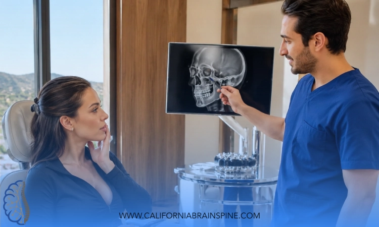
A brain MRI for dizziness is a scan generally preferred when a doctor suspects the dizziness comes from the brain itself rather than the inner ear. Radiology references describe MRI of the brain with diffusion-weighted imaging as the imaging choice for suspected brain causes, including reduced blood flow at the back of the brain, demyelinating disease (conditions that damage the protective coating around nerve fibers), tumors and inflammatory conditions.
The Importance of MRI in Dizziness Diagnosis
The importance of MRI in dizziness diagnosis comes from what it can see that a CT may not. A systematic review of emergency dizziness cases found that MRI outperformed CT for diagnosing acute dizziness, particularly for strokes in the posterior fossa, the region at the back of the brain that handles balance and coordination. When a scan is needed for a possible stroke, MRI is generally the more informative choice.
That still does not make MRI a routine first step for every dizzy person. Its value depends on the question being asked, and for many inner-ear problems that question can be answered at the bedside, as the sections below explain.
MRI for Vertigo: What It Can and Cannot Show
An MRI for vertigo is often normal when the cause is in the inner ear. Most inner-ear (peripheral) vestibular disorders, including benign paroxysmal positional vertigo (BPPV) and vestibular neuritis, typically do not show up on routine imaging. A clear MRI and very real vertigo can therefore sit side by side: the scan looks at the brain's structure, and many inner-ear problems leave nothing visible for it to find.

Why Do I Need a Brain Scan for Dizziness?
A brain scan for dizziness is usually recommended because something in the history or exam raises the possibility of a cause in the brain, the nerves or the blood vessels that an exam alone cannot settle. Radiology references and emergency research point to a fairly consistent set of features:
- Sudden dizziness or vertigo that comes with a focal neurological sign (a problem limited to one specific part of the body or one function, such as weakness on one side), severe trouble walking, or a new headache
- Eye-movement findings that look central rather than inner-ear, or other focal findings on the exam, which predicted acute brain lesions in one emergency-department study
- Dizziness alongside hearing loss or ringing on one side
- Hearing loss that keeps progressing, or a problem with one of the cranial nerves
- Vertigo that keeps coming back without an explanation, a suspected growth, a recent head injury, or a possible infection
The one-sided hearing feature deserves a word of its own. Research on hearing loss and imaging has found that people with uneven hearing loss together with ringing in one ear or dizziness are more likely to have an abnormal MRI finding, which is why this combination is one of the situations where an MRI is often considered.
If a doctor has recommended a scan, it often means one of these features showed up. If you were told you did not need one, it often means the exam fit a recognizable inner-ear pattern. Either way, you are entitled to ask which finding led to the decision.
Does Dizziness After a Head Injury Need an MRI?
Dizziness after a head injury may need imaging, because trauma is one of the recognized reasons a scan is considered. Radiology references list trauma among the imaging indications for dizziness, alongside the other features listed above.
Later on, the question shifts. If dizziness after a concussion is steady or slowly easing, the useful question is usually which part of the balance system is not coordinating well. If it is getting worse, or something new appears, such as vision changes, weakness, trouble speaking or a different kind of headache, it needs medical attention again, even if an earlier scan was clear.
An exam tests how the eyes, the inner ear and balance are working, and vestibular and neurological rehabilitation may be considered when clinically appropriate after evaluation.

Is a CT Scan for Dizziness Enough?
A CT scan for dizziness has a narrower role than many people expect, and a normal CT does not rule out a stroke in the balance areas at the back of the brain. CT has low sensitivity for an early stroke, especially in the posterior fossa, and the authors of an emergency-department study warned that a negative CT may falsely reassure a doctor that the cause sits in the inner ear.
The same study looked at how often CT found anything at all. In dizzy emergency patients who had not been pre-selected, brain CT found a cause in only about 2% of cases. A low yield is not the same as no value, but it explains why a CT is rarely the last word on dizziness.
When a scan is needed for a possible stroke, MRI is generally more informative than CT, which is one reason a doctor may still recommend an MRI after a normal CT.
Not Sure Whether Your Dizziness Needs a Scan or an Exam?
A 120-minute neurodiagnostic assessment in Calabasas looks at your eye movements, positional responses and balance, and makes clear when the next step belongs with medical neurology, ENT or your own physician.
Diagnosing Dizziness With Neurological Assessments: What Does the Exam Check?
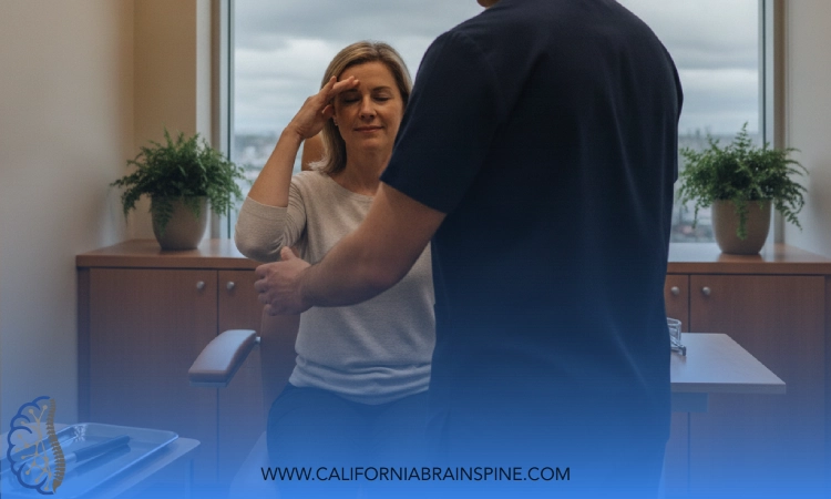
Diagnosing dizziness with neurological assessments means testing the systems that keep you balanced (the inner ear, the eyes and the brain's coordination of the two) before deciding whether a scan adds anything. Each part of the exam answers a different question.

Bedside Eye-Movement Testing (HINTS)
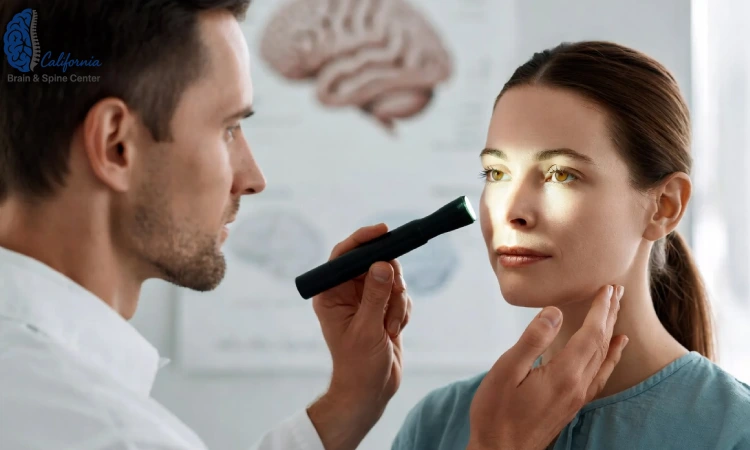
HINTS is a three-part bedside eye-movement exam used when dizziness starts suddenly and stays continuous, a picture called acute vestibular syndrome. In the same emergency-department study, no patient with acute vestibular syndrome whose complete HINTS exam was negative had an acute brain lesion, and the authors concluded that imaging may be dispensable in that situation. The study also found that central eye-movement signs and focal abnormalities on the neurological exam were associated with a brain cause.
Positional Testing (Dix-Hallpike)
The Dix-Hallpike maneuver is a positional test done on the exam table. Under the BPPV clinical practice guideline, posterior canal BPPV is diagnosed when this maneuver provokes vertigo together with a characteristic twisting, up-beating jerk of the eyes called nystagmus. No scan is part of that diagnosis.
Gait, Balance and Eye Tracking
A full evaluation also watches how you stand, walk and turn, how your eyes follow a moving target, and how they hold steady when your head moves. At California Brain & Spine Center, Dr. Alireza Chizari, DC, DACNB uses objective testing that may include videonystagmography (VNG), which records eye movements on video, and computerized assessment of postural stability, which measures sway while you stand. These tests describe how the balance system is working. Brain structure is the job of imaging, which is why the two answer different questions.
Side by side, the main tools compare like this:
| Tool | What it looks at | What it can tell you | What it cannot tell you |
|---|---|---|---|
| Brain MRI with diffusion-weighted imaging | The structure of the brain, including small areas of reduced blood flow | Generally preferred when a brain cause is suspected, and more informative than CT for strokes at the back of the brain | Most inner-ear causes, such as BPPV and vestibular neuritis, typically look normal on routine imaging |
| CT scan of the head | The structure of the brain | Found a cause in about 2% of unselected dizzy emergency patients in one study | A normal CT does not rule out an early stroke at the back of the brain and can falsely reassure |
| HINTS bedside eye-movement exam | Eye movements in sudden, continuous dizziness | In one study, no patient with a complete negative HINTS exam had an acute brain lesion; central signs point toward imaging | It does not show brain structure, so a central or unclear result leads to imaging |
| Dix-Hallpike positional test | How the eyes respond when the head changes position | Diagnoses typical posterior canal BPPV without a scan | Signs that do not fit BPPV call for further evaluation, which may include imaging |
Which Kinds of Dizziness Usually Do Not Need a Scan?
Dizziness that fits a recognizable inner-ear pattern on the exam often does not need a scan, and for typical BPPV the clinical practice guideline advises against imaging. The BPPV guideline states that clinicians should not obtain imaging in a patient who meets the diagnostic criteria for BPPV, unless there are other signs or symptoms that do not fit.
Spinning brought on by changes in head position is the pattern the positional test is designed to reproduce. When the test reproduces it in the typical way, the diagnosis is made in the exam room.
Inner-ear weakness after a condition such as vestibular neuritis is another example. Routine imaging is typically normal, and the next step is often rehabilitation. A clinical practice guideline on vestibular rehabilitation recommends, based on strong evidence, offering vestibular rehabilitation to people with one-sided or two-sided vestibular hypofunction (inner-ear balance weakness) who have impairments and functional limits from it.
Skipping a scan in these cases is a judgment about where the problem sits. The dizziness is still real, and it still deserves a plan.

When Is Dizziness an Emergency?
Dizziness is an emergency when it comes on suddenly with other stroke signs, such as trouble walking, vision trouble, one-sided weakness or numbness, confusion, trouble speaking or a sudden severe headache. The CDC lists sudden trouble walking, dizziness, loss of balance or lack of coordination among the signs of stroke. In that situation the question is never whether to book an MRI or a clinic visit.
When dizziness needs emergency care
Call 911 if dizziness arrives suddenly together with any of these signs of stroke:
- Sudden dizziness with trouble walking, loss of balance or poor coordination
- Sudden trouble seeing in one or both eyes
- Sudden numbness or weakness of the face, arm or leg, especially on one side
- Sudden confusion or trouble speaking
- A sudden severe headache
If symptoms come on suddenly or severely - a sudden severe headache, weakness, trouble speaking, vision loss, or fainting - that is an emergency. Call 911 or go to an emergency room, not a clinic appointment.
My CT Scan Was Normal and I Am Still Dizzy. What Now?
A normal CT scan with ongoing dizziness means the scan did not show the cause, and the next step is often a closer exam of the inner ear, eye movements and balance. If a brain cause is still suspected, MRI is generally more informative than CT, so your physician may recommend one.
Most inner-ear causes typically do not show up on routine imaging, so a clear scan and real dizziness can easily go together. I wrote a separate guide for the moment the MRI comes back clean as well: what a normal MRI does not rule out covers that question in depth.
Some patterns still point toward the brain after a clear scan, and they are worth knowing. Our article on early warning signs of central vestibular disorders describes the eye-movement changes, gait patterns and symptom clusters that prompt a closer look at central pathways.
What Does a Dizziness Diagnosis at a Brain Health Center Involve?
A dizziness diagnosis at a brain health center such as California Brain & Spine Center starts with a detailed history and a 120-minute neurodiagnostic assessment, before any decision about imaging is made. The assessment looks at eye movements, positional responses, balance and autonomic responses such as heart-rate variability.
One question patients often ask me is whether they should get an MRI before they come in. My answer depends on how the dizziness started. If it began suddenly with any of the warning signs above, that belongs in an emergency room today. If it has been building or recurring for weeks, the exam is a sensible place to start, because it can help show which part of the balance system is struggling and whether anything points toward the brain.
Another thing people often misunderstand is who does what. When something on the exam points toward a structural or vascular cause, the next step is a referral to medical neurology, ENT or your primary care physician, who decide on imaging and interpret it. Functional neurology works alongside them: the scan answers whether something is structurally wrong, and the exam describes how the balance system is functioning.
If the exam fits an inner-ear or functional pattern, care may include, when clinically appropriate after evaluation, vestibular and neurological rehabilitation and the other non-invasive services described on our services page.
Where Can I Have Dizziness Evaluated in Calabasas?
Dizziness evaluations with Dr. Alireza Chizari, DC, DACNB take place at California Brain & Spine Center on Park Granada in Calabasas, along the 101 corridor and a short drive from Woodland Hills. The office serves the West Valley and greater Los Angeles, for anyone whose dizziness does not fit a simple pattern and who wants a clear answer about the next step, whether that is rehabilitation, a referral to medical neurology or ENT, or emergency care.
Questions About MRI for Dizziness
These are the questions about MRI for dizziness I hear most often, answered briefly.
Should I Get an MRI for Headaches and Dizziness?
An MRI for headaches and dizziness is typically considered when the headache is new, or when it comes with severe trouble walking or other neurological signs. A sudden severe headache with dizziness is different: it can be a stroke sign, so call 911 instead. For long-standing, familiar headaches with dizziness, an exam is often the first step.
Is a Brain MRI for Dizziness Better Than a CT Scan?
For a possible stroke, a brain MRI for dizziness is generally more informative than a CT scan, and a systematic review found MRI outperformed CT for strokes at the back of the brain. A CT scan for dizziness still has a role, but a normal CT does not rule out a stroke in the balance areas.
Does an MRI for Vertigo Show BPPV?
Usually not. An MRI for vertigo caused by BPPV is typically normal, because most inner-ear causes do not show up on routine imaging. Typical BPPV is diagnosed with a positional test, the Dix-Hallpike maneuver, and the clinical guideline advises against imaging a person who meets the criteria unless other signs do not fit.
Can I Have Dizziness Evaluated in Calabasas Without a Scan First?
Often, yes. If your dizziness has been building or recurring rather than starting suddenly with stroke signs, a 120-minute neurodiagnostic assessment in Calabasas can look at eye movements, positional responses and balance first. If anything points toward a brain cause, the next step is a referral to medical neurology or your physician, who decide on an MRI for dizziness.
My MRI Was Normal. Can My Dizziness Still Be Evaluated?
Yes. A normal MRI for dizziness answers whether a structural brain cause is visible, and most inner-ear problems typically look normal on routine imaging. An exam of eye movements, positional responses, gait and balance can still help show which part of the balance system is involved and whether vestibular rehabilitation may be appropriate.
If you are weighing an MRI for dizziness, the deciding factor is usually what the history and exam show. Sudden dizziness with stroke signs belongs in an emergency room. Dizziness that has been building, recurring, or lingering after a scan came back clear deserves a careful look at the inner ear, the eyes and balance, along with a straight answer about whether imaging is the next step. That is the work I do every day in Calabasas, and I am glad to help you sort out which path fits.
California Brain & Spine Center | (818) 649-5300 | Calabasas, CA 91302. Book online at californiabrainspine.janeapp.com.
This content is for educational purposes only and is not medical advice. Consult a qualified healthcare provider about your specific situation.
Get a Clear Answer About Your Dizziness
Dr. Chizari's assessment is designed to show how your eyes, inner ear and balance are working together, and to explain whether rehabilitation, a referral or emergency care fits your situation.