Antisaccades in Parkinson’s Disease: What Eye Movements Reveal About Motor Control
Parkinson’s disease is often recognized by its visible motor symptoms—tremor, stiffness, slowed movement, shuffling gait, or changes in balance. But long before movement becomes obviously impaired, the brain’s motor control systems may begin showing subtle signs of dysfunction.
One of the most fascinating ways clinicians and researchers study these systems is through eye movements.
More specifically, antisaccades a type of controlled eye movement that requires the brain to suppress an automatic response—can provide valuable insight into how well the brain manages motor planning, inhibition, attention, and executive control.
At California Brain & Spine Center, led by Dr. Alireza Chizari, DC, DACNB, advanced functional neurological and functional assessments may include careful evaluation of visual tracking, eye movement control, balance, vestibular function, and brain-body coordination. For patients with Parkinson’s disease or Parkinsonian symptoms, understanding how the eyes move can help reveal important information about the nervous system’s ability to regulate movement.
This article explores the role of antisaccades in Parkinson’s disease, what abnormal eye movements may indicate, and why eye movement testing can be a meaningful window into motor control and brain health.
What Are Antisaccades?
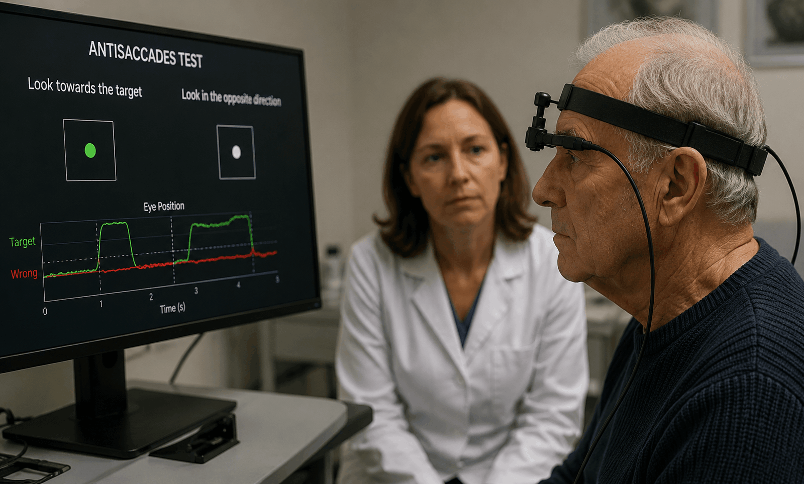
A saccade is a quick eye movement that shifts your gaze from one point to another. For example, if something suddenly appears on your right side, your eyes naturally and automatically move toward it. This is called a prosaccade.
An antisaccade is different.
During an antisaccade task, a person must look in the opposite direction of a visual target. If a stimulus appears on the right, the person must suppress the automatic urge to look right and instead look left.
That sounds simple, but neurologically, it is a complex task.
To perform an antisaccade correctly, the brain must:
- Detect the visual stimulus
- Inhibit the automatic reflex to look toward it
- Plan an intentional eye movement in the opposite direction
- Execute that movement accurately
- Monitor and correct mistakes if needed
This requires coordination between several brain regions, including the frontal eye fields, prefrontal cortex, basal ganglia, superior colliculus, cerebellum, and brainstem eye movement centers.
Because Parkinson’s disease affects motor control networks—especially circuits involving the basal ganglia—antisaccade performance can reveal important clues about how the disease impacts movement regulation.
Why Eye Movements Matter in Parkinson’s Disease
Parkinson’s disease is not only a disorder of the hands, legs, or gait. It is a disorder of motor control across the nervous system.
Eye movements are motor actions too.
Although they are small and fast, eye movements depend on many of the same systems involved in larger body movements, including:
- Motor initiation
- Movement timing
- Inhibition of unwanted movements
- Voluntary movement planning
- Cognitive control
- Attention shifting
- Error correction
This is why eye movement testing can be so valuable. The eyes may reveal subtle problems in motor control before they become obvious in walking, balance, or hand coordination.
In Parkinson’s disease, patients may show changes in:
- Saccade speed
- Saccade accuracy
- Reaction time
- Difficulty suppressing automatic eye movements
- Increased antisaccade errors
- Delayed voluntary gaze shifts
- Reduced ability to correct eye movement mistakes
These findings can help clinicians better understand how Parkinson’s disease is affecting the brain’s control systems.
The Link Between Antisaccades and Motor Control
Motor control is not simply about moving. It is about moving at the right time, in the right direction, with the right amount of force, while suppressing unnecessary or competing movements.
Antisaccades test this process in a precise way.
When a person performs an antisaccade, the brain must stop one movement plan and generate another. This is similar to many everyday motor tasks, such as:
- Stopping before stepping into traffic
- Changing direction while walking
- Avoiding an obstacle
- Reaching for the correct object
- Adjusting posture when balance changes
- Suppressing an impulsive movement
In Parkinson’s disease, the basal ganglia and related motor circuits become less efficient at regulating movement initiation and inhibition. This may contribute to symptoms such as:
- Bradykinesia, or slowness of movement
- Freezing episodes
- Delayed reaction time
- Difficulty starting movement
- Difficulty stopping or switching movement
- Reduced automatic movement control
- Impaired balance adjustments
Antisaccade testing gives clinicians a way to observe similar control problems through fast, measurable eye movements.
Antisaccade Errors in Parkinson’s Disease
One of the most common findings in Parkinson’s disease research is an increase in antisaccade errors.
An antisaccade error occurs when a person looks toward the stimulus instead of away from it. In other words, the automatic response wins before the brain can suppress it.
This may suggest difficulty with:
- Response inhibition
- Executive control
- Motor suppression
- Attention regulation
- Frontal-basal ganglia communication
- Cognitive flexibility
In Parkinson’s disease, these errors may be especially meaningful because they can reflect more than just eye movement dysfunction. They may indicate reduced efficiency in the brain networks responsible for controlling voluntary movement and suppressing inappropriate motor responses.
Delayed Antisaccades and Slowed Movement Initiation
Another important finding is delayed antisaccade reaction time.
People with Parkinson’s disease may take longer to initiate voluntary eye movements, especially when the task requires inhibition and conscious control. This delay may parallel the broader symptom of bradykinesia, or slowed movement.
A delayed antisaccade may suggest that the brain is taking longer to:
- Process the instruction
- Suppress the automatic eye movement
- Create the voluntary movement plan
- Send the correct motor command
- Execute the movement accurately
This can be especially relevant in patients who report:
- Slower thinking
- Delayed reactions
- Trouble multitasking
- Difficulty initiating movement
- Problems with coordination
- Reduced confidence while walking or turning
Because eye movements are fast and measurable, they may help reveal subtle timing problems in the nervous system.
The Role of the Basal Ganglia in Antisaccades
The basal ganglia are deep brain structures heavily involved in movement control, habit formation, motor selection, and inhibition. Parkinson’s disease is strongly associated with dysfunction in basal ganglia circuits, particularly due to dopamine loss.
These circuits help the brain decide which movements to allow and which movements to suppress.
In an antisaccade task, the basal ganglia help regulate whether the automatic eye movement toward the stimulus should be blocked so that the correct opposite movement can occur.
When basal ganglia function is disrupted, the person may be more likely to:
- Look toward the target by mistake
- React too slowly
- Have trouble switching gaze direction
- Show inconsistent eye movement accuracy
- Struggle with voluntary motor control
This is why antisaccades can be useful for studying Parkinson’s disease. They engage the same brain systems that are often impaired in the condition.
Antisaccades, Executive Function, and Parkinson’s Disease
Parkinson’s disease can affect more than movement. Many patients also experience changes in executive function, including:
- Slower mental processing
- Reduced working memory
- Difficulty multitasking
- Trouble shifting attention
- Impaired planning
- Reduced impulse control
- Difficulty adapting to changing situations
Antisaccade performance depends heavily on executive control. The person must remember the rule, resist an automatic response, and generate the correct intentional response.
For this reason, antisaccade errors may reflect both motor and cognitive changes.
In some patients, increased antisaccade errors may be associated with:
- Mild cognitive impairment
- Reduced attention control
- Frontal lobe dysfunction
- Slower decision-making
- Difficulty with dual-task activities
- Greater challenges in daily functioning
This does not mean that antisaccade testing alone can diagnose cognitive decline. However, it may provide valuable information when combined with a comprehensive neurological evaluation.
Eye Movements and Parkinsonian Gait or Balance Problems
Many people with Parkinson’s disease experience gait and balance difficulties, including:
- Shortened steps
- Shuffling gait
- Freezing of gait
- Poor turning control
- Postural instability
- Increased fall risk
- Difficulty walking while looking around
Eye movement control is closely related to balance and navigation. The eyes help guide the body through space. Before we step, turn, or reach, the eyes often move first to gather information.
If gaze control is impaired, the brain may have less accurate visual input for movement planning.
In Parkinson’s disease, abnormal eye movement patterns may contribute to:
- Poor spatial orientation
- Delayed obstacle detection
- Difficulty scanning the environment
- Reduced confidence while walking
- Increased instability during turns
- Trouble coordinating head, eye, and body movement
This is one reason a functional neurological assessment may include eye movement, vestibular, and balance testing together.
Can Antisaccade Testing Help Detect Early Parkinson’s Changes?
Antisaccade abnormalities are not exclusive to Parkinson’s disease. They can occur in many neurological and cognitive conditions, including concussion, traumatic brain injury, cerebellar dysfunction, vestibular disorders, neurodegenerative conditions, and executive function disorders.
However, in Parkinson’s disease, antisaccade testing may help identify subtle dysfunction in motor control networks.
Potentially relevant findings may include:
- Increased direction errors
- Delayed voluntary eye movements
- Reduced saccade amplitude
- Poor gaze accuracy
- Difficulty correcting mistakes
- Impaired response inhibition
- Inconsistent performance under cognitive load
These findings do not replace medical diagnosis. Parkinson’s disease diagnosis requires a clinical examination, medical history, neurological assessment, and sometimes imaging or medication response evaluation.
But antisaccade testing can be a useful part of a broader picture.
Antisaccades and Medication Effects in Parkinson’s Disease
Dopamine plays a major role in Parkinson’s disease, and dopamine-based medications can affect motor performance.
Some studies suggest that eye movement patterns may change depending on medication state. For example, certain patients may perform differently when they are in an “on” medication state compared with an “off” state.
Eye movement testing may help clinicians observe changes in:
- Reaction time
- Movement initiation
- Motor inhibition
- Accuracy
- Fatigue effects
- Cognitive-motor performance
However, medication effects can vary significantly between patients. Antisaccade testing should be interpreted carefully and always in the context of the patient’s full clinical picture.
What Abnormal Antisaccades May Reveal About Brain Function
Abnormal antisaccades in Parkinson’s disease may suggest that the brain is having difficulty coordinating several important processes at once.
These include:
1. Response Inhibition
The ability to stop an automatic or unwanted response.
2. Voluntary Motor Planning
The ability to intentionally generate a movement according to a goal or instruction.
3. Cognitive Flexibility
The ability to shift from one response pattern to another.
4. Attention Control
The ability to focus on the task and resist distraction.
5. Error Monitoring
The ability to recognize and correct mistakes quickly.
6. Basal Ganglia-Frontal Communication
The coordination between deep motor circuits and frontal executive control regions.
In Parkinson’s disease, these systems may become less efficient, which can affect both eye movements and whole-body movement.
How California Brain & Spine Center Evaluates Eye Movement and Motor Control
At California Brain & Spine Center, patients with Parkinson’s disease, Parkinsonian symptoms, balance problems, dizziness, post-concussion symptoms, or unexplained functional neurological complaints may benefit from a detailed functional assessment.
Depending on the patient’s symptoms and clinical needs, evaluation may include:
- functional Neurological history and symptom review
- Eye movement assessment
- Visual tracking evaluation
- Saccade and antisaccade observations
- Balance and postural control testing
- Vestibular screening
- Gait and coordination assessment
- Cognitive-motor function review
- Review of prior imaging or medical records when relevant
The goal is not simply to identify symptoms, but to understand how the nervous system is functioning as a whole.
For Parkinson’s disease patients, this may help clarify how motor control, visual guidance, balance, and executive function are interacting.
Can Eye Movement Exercises Help Parkinson’s Disease?
Eye movement exercises do not cure Parkinson’s disease. Parkinson’s is a progressive functional neurological condition that requires medical management and ongoing care.
However, targeted neurological rehabilitation may support certain functional goals in some patients.
Depending on the individual, therapy may focus on:
- Visual tracking
- Gaze stabilization
- Saccade control
- Antisaccade training
- Balance integration
- Vestibular coordination
- Reaction time
- Postural awareness
- Cognitive-motor tasks
- Gait and visual scanning strategies
The purpose of these exercises is to challenge and support the brain’s ability to coordinate sensory input and motor output.
For some patients, improving visual-motor control may support better balance, navigation, attention, and confidence during movement. However, any exercise program should be individualized and supervised by a qualified clinician.
Antisaccades vs. Other Eye Movement Tests in Parkinson’s Disease
Antisaccades are only one type of eye movement assessment. Other eye movement tests may also provide useful information.
These may include:
Prosaccades
Looking directly toward a target. This helps evaluate basic reflexive gaze shifting.
Smooth Pursuits
Following a moving target smoothly with the eyes. This helps assess visual tracking and coordination.
Fixation Testing
Holding the eyes steady on a target. This may reveal instability or unwanted eye movements.
Gaze Holding
Maintaining gaze in different directions. This can help assess brainstem and cerebellar function.
Vestibulo-Ocular Reflex Testing
Evaluating how the eyes stabilize when the head moves. This is especially important for dizziness and balance problems.
Together, these tests can provide a broader understanding of brain, vestibular, and motor function.
Case Example: Subtle Eye Movement Changes in a Patient With Parkinsonian Symptoms
A patient with early Parkinsonian symptoms visited California Brain & Spine Center reporting slower movements, mild balance issues, and difficulty turning while walking. The patient also noticed that busy environments felt overwhelming and that they had trouble quickly shifting attention.
During functional assessment, the patient demonstrated delayed voluntary eye movements and increased difficulty with tasks requiring suppression of automatic gaze responses. Antisaccade-style testing showed that the patient sometimes looked toward a stimulus instead of away from it, especially when fatigued.
These findings suggested challenges with response inhibition, visual-motor timing, and cognitive-motor coordination.
The care plan included individualized functional neurological rehabilitation focused on gaze control, balance integration, visual scanning, and movement timing. Over time, the patient reported improved confidence during walking and better awareness when moving through visually complex environments.
This example is for educational purposes only and does not represent a guaranteed outcome. Each patient’s condition, diagnosis, and response to care are unique.
When Should Someone With Parkinson’s Consider Eye Movement Evaluation?
A person with Parkinson’s disease or suspected Parkinsonian symptoms may benefit from eye movement and motor control evaluation if they experience:
- Balance problems
- Dizziness or visual disorientation
- Difficulty walking in busy environments
- Freezing or hesitation with movement
- Increased falls or near-falls
- Trouble turning
- Slowed reaction time
- Brain fog or cognitive slowing
- Difficulty multitasking
- Poor coordination
- Visual tracking problems
- Trouble shifting gaze
- Post-concussion symptoms alongside Parkinsonian features
Eye movement testing may be especially useful when symptoms are complex and involve both movement and cognition.
Why Antisaccades Are a Window Into Brain Health
Antisaccades are powerful because they are simple to perform but functional neurologically rich.
They reveal how well the brain can:
- Stop an automatic response
- Create a voluntary movement
- Coordinate vision and action
- Use attention effectively
- Control impulses
- Monitor performance
- Adapt to changing instructions
In Parkinson’s disease, these abilities are deeply connected to the same systems involved in motor control. That is why abnormal antisaccades may offer insight into the broader functional neurological challenges patients face.
They are not just eye movements. They are a reflection of brain-body coordination.
Frequently Asked Questions
What are antisaccades in Parkinson’s disease?
Antisaccades are intentional eye movements made in the opposite direction of a visual target. In Parkinson’s disease, antisaccade testing can help evaluate motor control, response inhibition, attention, and executive function.
Why do people with Parkinson’s make antisaccade errors?
People with Parkinson’s may make antisaccade errors because the brain has difficulty suppressing automatic responses. This may relate to changes in basal ganglia and frontal brain circuits involved in motor inhibition and executive control.
Can antisaccade testing diagnose Parkinson’s disease?
No. Antisaccade testing alone cannot diagnose Parkinson’s disease. Diagnosis requires a full medical and neurological evaluation. However, antisaccade testing may provide useful information about motor control and brain function.
What do delayed eye movements mean in Parkinson’s disease?
Delayed eye movements may suggest slowed voluntary motor initiation, impaired processing speed, or difficulty coordinating visual and motor systems. In Parkinson’s disease, this may relate to broader movement slowing or cognitive-motor dysfunction.
Are eye movement problems common in Parkinson’s disease?
Yes, many people with Parkinson’s disease may experience changes in saccades, visual tracking, gaze control, reaction time, and eye movement accuracy. These changes can vary from person to person.
Can eye movement exercises improve Parkinson’s symptoms?
Eye movement exercises do not cure Parkinson’s disease. However, targeted visual-motor and neurological rehabilitation may help some patients improve gaze control, balance integration, visual scanning, and movement confidence.
How are antisaccades related to executive function?
Antisaccades require the brain to suppress an automatic response and generate an intentional movement. This depends on executive functions such as inhibition, attention, working memory, and cognitive flexibility.
Why are the basal ganglia important for antisaccades?
The basal ganglia help regulate movement selection and inhibition. Since Parkinson’s disease affects basal ganglia circuits, antisaccade performance may reveal problems with suppressing incorrect movements and initiating correct ones.
Should Parkinson’s patients get eye movement testing?
Patients with Parkinson’s who have balance problems, visual disorientation, slowed reactions, gait issues, or cognitive-motor symptoms may benefit from a comprehensive evaluation that includes eye movement testing.
Where can I get eye movement and motor control evaluation in California?
California Brain & Spine Center in Calabasas, California, provides neurological and functional assessments that may include eye movement, balance, vestibular, and motor control evaluation.
Conclusion
Parkinson’s disease affects far more than visible body movement. It can influence the brain’s ability to plan, inhibit, initiate, and coordinate movement across multiple systems—including the eyes.
Antisaccades in Parkinson’s disease offer a valuable way to understand these changes. When a person struggles to look away from a target, reacts slowly, or makes repeated gaze errors, it may reveal deeper challenges in motor control, executive function, attention, and basal ganglia-frontal communication.
While antisaccade testing does not diagnose Parkinson’s disease by itself, it can be an important part of a comprehensive functional neurological evaluation. By studying eye movements, clinicians can gain a clearer picture of how the brain controls action, suppresses unwanted responses, and coordinates movement in daily life.
For patients with Parkinson’s disease, Parkinsonian symptoms, balance problems, dizziness, or cognitive-motor difficulties, a detailed eye movement and neurological assessment may help guide a more personalized care plan.
If you or a loved one is experiencing Parkinson’s disease symptoms, changes in balance, slowed movement, visual tracking problems, dizziness, or difficulty with coordination, the team at California Brain & Spine Center can help evaluate how your brain and body are working together.
Schedule a consultation with Dr. Alireza Chizari, DC, DACNB and the California Brain & Spine Center team to learn more about functional neurological assessment, eye movement evaluation, and personalized care options.
Comments
FAQ
What is Functional Neurology?
Functional Neurology is a healthcare specialty that focuses on assessing and rehabilitating the nervous system’s function. It emphasizes neuroplasticity—the brain’s ability to adapt and reorganize—using non-invasive, evidence-based interventions to improve neurological performance.
How does Functional Neurology differ from traditional neurology?
Traditional neurology often concentrates on diagnosing and treating neurological diseases through medications or surgery. In contrast, Functional Neurology aims to optimize the nervous system’s function by identifying and addressing dysfunctions through personalized, non-pharmaceutical interventions.
Is Functional Neurology a replacement for traditional medical care?
No. Functional Neurology is intended to complement, not replace, traditional medical care. Practitioners often collaborate with medical professionals to provide comprehensive care.
What conditions can Functional Neurology help manage?
Functional Neurology has been applied to various conditions, including:
• Concussions and Post-Concussion Syndrome
• Traumatic Brain Injuries (TBI)
• Vestibular Disorders
• Migraines and Headaches
• Neurodevelopmental Disorders (e.g., ADHD, Autism)
• Movement Disorders
• Dysautonomia
• Peripheral Neuropathy
• Functional Neurological Disorder (FND)
Can Functional Neurology assist with neurodegenerative diseases?
While Functional Neurology does not cure neurodegenerative diseases, it can help manage symptoms and improve quality of life by optimizing the function of existing neural pathways.
What diagnostic methods are used in Functional Neurology?
Functional Neurologists employ various assessments, including:
• Videonystagmography (VNG)
• Computerized Posturography
• Oculomotor Testing
• Vestibular Function Tests
• Neurocognitive Evaluations
How is a patient’s progress monitored?
Progress is tracked through repeated assessments, patient-reported outcomes, and objective measures such as balance tests, eye movement tracking, and cognitive performance evaluations.
What therapies are commonly used in Functional Neurology?
Interventions may include:
- Vestibular Rehabilitation
- Oculomotor Exercises
- Sensorimotor Integration
- Cognitive Training
- Balance and Coordination Exercises
- Nutritional Counseling
- Lifestyle Modifications
Are these therapies personalized?
Absolutely. Treatment plans are tailored to the individual’s specific neurological findings, symptoms, and functional goals.
Who can benefit from Functional Neurology?
Individuals with unresolved neurological symptoms, those seeking non-pharmaceutical interventions, or patients aiming to optimize brain function can benefit from Functional Neurology.
Is Functional Neurology suitable for children?
Yes. Children with developmental delays, learning difficulties, or neurodevelopmental disorders may benefit from Functional Neurology approaches.
How does Functional Neurology complement other medical treatments?
It can serve as an adjunct to traditional medical care, enhancing outcomes by addressing functional aspects of the nervous system that may not be targeted by conventional treatments.
How is technology integrated into Functional Neurology?
Technological tools such as virtual reality, neurofeedback, and advanced diagnostic equipment are increasingly used to assess and enhance neurological function.
What is the role of research in Functional Neurology?
Ongoing research continues to refine assessment techniques, therapeutic interventions, and our understanding of neuroplasticity, contributing to the evolution of Functional Neurology practices.

Dr. Alireza Chizari
Latest articles