Antisaccades After Concussion: How Eye Movements Can Detect Subtle Brain Dysfunction
If you have had a concussion and you still feel “off,” even when standard imaging looks normal, I want you to know that your symptoms are real. Brain fog, dizziness, visual discomfort, poor focus, delayed reaction time, and feeling overwhelmed in busy environments can all reflect subtle changes in how the brain coordinates attention, balance, vision, and movement.
I, Dr. Alireza Chizari DACNB, will explain in this article how we evaluate antisaccades after concussion and why these small, fast eye movements can reveal important information about subtle brain dysfunction. Your eyes do more than help you see. They also give us a window into how your brain controls attention, inhibition, timing, and neurological coordination.
At California Brain & Spine Center in Calabasas, California, my goal is not to reduce your story to a single symptom or test result. You are the hero of this process. You are the one living with the symptoms, trying to return to work, school, driving, exercise, family life, and confidence. My role is to be the guide who helps you understand what may be happening and what can be done next.
This page is about how antisaccades after concussion may help uncover hidden neurological stress, how we use eye movement testing as part of a broader concussion evaluation, and how personalized care may support recovery through vestibular rehabilitation, cognitive rehabilitation, NeuroSensory Integration, neuroplasticity rehabilitation, and other non-invasive neurological therapies when appropriate.
Why Antisaccades After Concussion Matter More Than Most People Realize
After a concussion, many patients are told to rest, wait, and return slowly to activity. For some people, symptoms resolve quickly. But for others, the brain does not fully return to efficient function right away. They may pass basic neurological exams, have normal CT or MRI results, and still struggle with symptoms that affect daily life.
This is where antisaccades after concussion become clinically meaningful. An antisaccade is a voluntary eye movement in which you are asked to look away from a sudden visual target instead of looking toward it. That sounds simple, but it requires the brain to perform several tasks at once:
It must notice the stimulus, suppress the automatic urge to look toward it, calculate the opposite direction, and then generate a controlled eye movement. This process involves attention, impulse control, frontal lobe function, basal ganglia circuits, cerebellar timing, brainstem pathways, and visual-vestibular integration.
When someone has more antisaccade errors, delayed responses, poor accuracy, or inconsistent timing after a concussion, it may suggest that the brain is having difficulty regulating automatic responses. In other words, the eyes may reveal what the patient has already been feeling: “My brain is working harder than it should.”
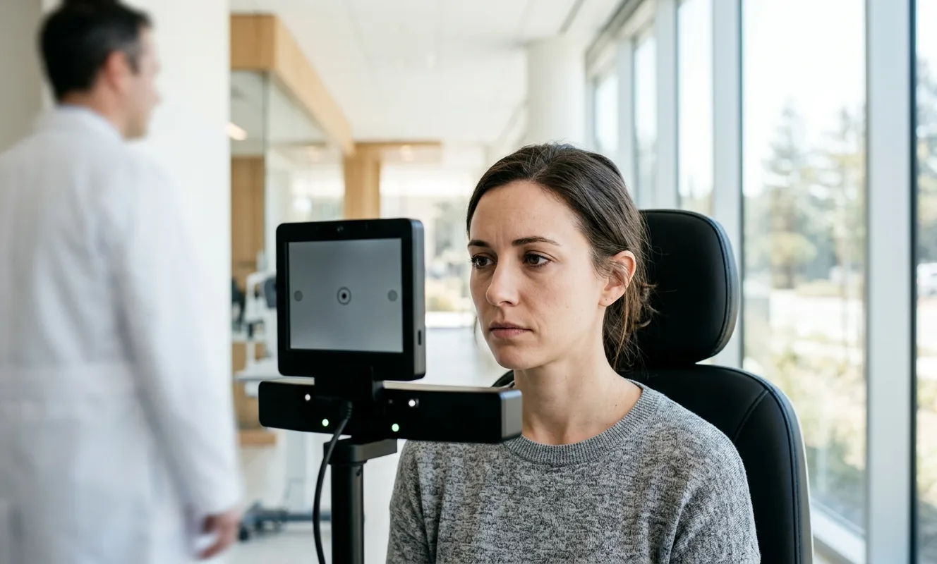
Image note: “A calm clinical scene showing a patient completing an eye movement test on a screen while a neurologically trained doctor observes in a modern Calabasas clinic, soft lighting, professional and reassuring atmosphere.”
The Hidden Brain Work Behind a Simple Eye Movement
When I evaluate a patient with post-concussion symptoms, I do not look at eye movements as isolated mechanical actions. I look at them as outputs of a complex neurological system. A small delay in an eye movement can sometimes reflect a much larger problem in brain communication.
The antisaccade task is especially valuable because it challenges executive control. Executive control is the brain’s ability to pause, choose, inhibit, organize, and respond appropriately. After concussion, patients often describe problems that sound very similar:
They may say, “I cannot focus like I used to,” “I lose my place when reading,” “My reaction time feels slower,” or “Busy stores make me dizzy and exhausted.” These symptoms can be connected to the same networks involved in antisaccade control.
Antisaccades after concussion are not just about the eyes. They are about the brain’s ability to regulate behavior under sensory load.
Healing often begins when you finally have a clear explanation for symptoms you were afraid no one could see.
What an Antisaccade Test Can Show After a Concussion
An antisaccade test can help reveal whether the brain is struggling with inhibition, accuracy, timing, or visual attention. It does not replace a full clinical evaluation, and it should not be used as a stand-alone diagnosis. But when interpreted correctly, it can provide important clues.
In my clinical approach, I consider how the patient performs, how they feel during testing, and whether the findings match their daily symptoms. For example, a patient who makes frequent errors on antisaccade testing may also report impulsive visual reactions, trouble scanning the environment, difficulty reading, or feeling overstimulated in visually busy places.
A patient with delayed antisaccades may describe slowed thinking, brain fog, difficulty multitasking, or mental fatigue after short periods of concentration. A patient with poor accuracy may have subtle problems with spatial processing, visual coordination, or cerebellar timing.
This is why antisaccades after concussion can be so useful. They help connect symptoms to function.
Antisaccade Errors and Response Inhibition
One of the most important findings we look for is whether the patient looks toward the target when they were supposed to look away. This is called a direction error. In many cases, this reflects difficulty suppressing an automatic response.
After concussion, the brain may become less efficient at filtering, inhibiting, and prioritizing information. This can show up as distractibility, poor focus, irritability, visual overwhelm, or reduced tolerance for complex environments.
In practical terms, the patient may not simply have an “eye problem.” They may have a brain control problem that appears through the eyes.
Delayed Antisaccades and Brain Fog
Some patients do not make many errors, but they respond slowly. Their eyes eventually move in the correct direction, but the timing is delayed. This can be clinically relevant because many concussion patients describe the same experience in daily life: they can do the task, but everything takes more effort and more time.
This is where antisaccades after concussion can support a deeper understanding of brain fog. Brain fog is not laziness, weakness, or lack of motivation. It can reflect measurable changes in processing speed, attention, sensory integration, autonomic regulation, and fatigue management.
Image note: “A visual concept image of the brain connected to the eyes with subtle glowing neural pathways, representing attention, inhibition, and concussion recovery, clean medical illustration style.”
Why Normal Imaging Does Not Always Mean Normal Function
Many patients feel confused when they are told their MRI or CT scan is normal, yet they still cannot function the way they did before the concussion. I always explain that structural imaging and functional performance are not the same thing.
A CT scan is excellent for detecting serious bleeding or fracture. An MRI can reveal certain structural injuries. But many concussion-related problems occur at the level of network function, sensory integration, timing, autonomic regulation, and metabolic stress. These changes may not appear on standard imaging.
That is why a functional neurological evaluation can be so important. Antisaccades after concussion may help show how efficiently the brain is controlling attention and movement in real time.
This does not mean every abnormal eye movement proves a concussion injury. Eye movements can be affected by sleep, medications, anxiety, migraine, vestibular disorders, neck injury, visual problems, and other neurological conditions. But when the pattern fits the history and symptoms, eye movement testing can become a valuable part of the clinical picture.
How I Approach Antisaccades After Concussion in Calabasas
At California Brain & Spine Center, I bring a unique background to this work. Before becoming a Doctor of Chiropractic and pursuing postdoctoral education in Clinical Neuroscience, I studied Electrical Engineering in Iran, completed a master’s degree in Advanced Engineering & Management in the UK, and worked in the United States as a Solar Engineer.
That engineering background shaped how I think. I look for patterns, systems, inputs, outputs, timing, feedback loops, and functional breakdowns. Later, my clinical neuroscience training helped me apply that systems-based thinking to the human nervous system.
When I evaluate antisaccades after concussion, I am not just asking, “Did the eyes move correctly?” I am asking, “What does this tell us about the patient’s brain, vestibular system, visual processing, autonomic state, and capacity to recover?”
A thoughtful concussion evaluation may include:
- ✅ Eye movement testing, including saccades, pursuits, fixation, convergence, and antisaccades
- ✅ Vestibular and balance assessment to understand dizziness, motion sensitivity, and spatial orientation
- ✅ Cognitive and sensory screening to evaluate brain fog, attention, fatigue, and visual overwhelm
- ✅ Review of symptoms, injury history, sleep, stress, neck involvement, and daily functional limitations
- ✅ Personalized recommendations for rehabilitation, therapy, or referral when needed
The goal is not to chase a test result. The goal is to understand the person in front of me.
You are not starting over. You are starting from a clearer understanding of what your brain needs next.
The Role of Eye Movements in Post-Concussion Dizziness and Visual Disturbance
At California Brain & Spine Center, patients are often evaluated for dizziness, balance problems, visual disturbances after concussion, brain fog, and memory concerns. Eye movements are closely connected to all of these symptoms because the visual system, vestibular system, neck proprioception, and brainstem must work together to keep the world stable.
When these systems are not properly integrated, patients may feel dizzy in grocery stores, uncomfortable while driving, nauseated when scrolling on a phone, or exhausted after reading. These symptoms can be frightening because they are often invisible to others.
Antisaccades after concussion provide one way to challenge the brain’s control system. They can help clinicians understand whether symptoms are related to poor inhibition, delayed processing, visual-vestibular mismatch, or fatigue under neurological demand.
Visual Disturbances After Concussion Are Not Always an Eye Exam Problem
A standard eye exam is important and can identify problems with refraction, eye health, or certain binocular vision issues. But many post-concussion visual symptoms are neurological. The eyes may be healthy, while the brain has difficulty coordinating visual input.
This is why some patients say, “My eye doctor said my eyes are fine, but I still cannot read comfortably.” In those cases, the issue may involve ocular motor control, vestibular integration, attention, or sensory processing.
How Eye Movement Testing Supports Personalized Rehabilitation
Eye movement findings help guide care. A patient with poor visual tracking may need a different approach than a patient with strong tracking but poor inhibition. A patient with dizziness triggered by head movement may need vestibular rehabilitation. A patient with brain fog and fatigue may need carefully paced cognitive rehabilitation and neuroplasticity-based exercises.
At California Brain & Spine Center, care may include vestibular rehabilitation, cognitive rehabilitation, NeuroSensory Integration, neuroplasticity rehabilitation, and non-invasive functional neurology therapy when appropriate. Tools such as Low-Level Laser Therapy, PEMF, HBOT, GammaCore Vagus Nerve Stimulation, and the NeuroRevive Program may be considered depending on the patient’s presentation, safety factors, and clinical goals.
Image note: “A professional rehabilitation setting with a patient doing guided visual-vestibular exercises, a doctor nearby monitoring progress, calm clinical environment, hopeful and focused mood.”
What Subtle Brain Dysfunction Can Feel Like After Concussion
Subtle brain dysfunction does not always feel subtle to the patient. It can affect work, school, parenting, exercise, relationships, and emotional confidence. The person may look normal from the outside but feel unstable on the inside.
Common experiences may include visual fatigue, headaches, dizziness, difficulty concentrating, slower thinking, memory lapses, anxiety in busy spaces, motion sensitivity, poor sleep, and reduced tolerance for screens. These symptoms can fluctuate. A patient may feel decent in the morning and overwhelmed by the afternoon.
This is why antisaccades after concussion should be interpreted with the full story in mind. A single test does not define a person. But the right test, used at the right time, can help explain why the brain is struggling.
How California Brain & Spine Center Builds a Recovery Plan
At California Brain & Spine Center in Calabasas, the evaluation process is designed to understand the patient’s neurological patterns, not just label the condition. Patients from Southern California and beyond seek care when symptoms persist, when standard answers feel incomplete, or when they need a more detailed functional approach.
The clinic’s approach is evidence-informed, personalized, and non-invasive whenever possible. The team considers how concussion may affect the visual system, vestibular system, autonomic nervous system, cognitive performance, sleep regulation, neck function, and emotional resilience.
A personalized care plan may focus on:
- ✨ Improving visual tracking and eye movement control
- ✨ Reducing dizziness, motion sensitivity, and visual overwhelm
- ✨ Supporting attention, memory, and cognitive endurance
- ✨ Rebuilding balance, coordination, and confidence in movement
- ✨ Helping the brain adapt through neuroplasticity-based rehabilitation
The NeuroRevive Program may be used as part of a broader strategy to support brain injury recovery, depending on the patient’s needs. Care is always adapted to tolerance. After concussion, pushing too hard can flare symptoms. But doing too little can also slow progress. The art is finding the right level of challenge.
Recovery is not about forcing the brain to perform. It is about giving the brain the right signal, at the right intensity, at the right time.
Antisaccades After Concussion and Return to Work, School, and Driving
One reason antisaccades after concussion matter is that they can relate to real-life performance. The ability to suppress distractions, shift attention, react appropriately, and process visual information is essential for daily activities.
Returning to work may require screen tolerance, multitasking, reading, visual scanning, and sustained attention. Returning to school may require note taking, switching focus from board to paper, reading under time pressure, and learning in visually busy classrooms. Driving requires fast visual processing, peripheral awareness, eye-head coordination, inhibition, and reaction timing.
If the brain is still inefficient after concussion, these tasks can feel exhausting or unsafe. Eye movement testing can help identify why a patient feels overloaded and what types of rehabilitation may support safer progression.
Why Symptoms Can Flare in Busy Environments
Many patients feel worse in grocery stores, shopping malls, airports, restaurants, traffic, or crowded hallways. These environments create high visual and sensory demand. The brain must filter motion, sound, light, balance input, and spatial information all at once.
If antisaccade performance is poor, it may reflect difficulty inhibiting irrelevant visual stimuli. This can help explain why a patient feels pulled in every direction by the environment.
Why Reading and Screen Use Can Become Difficult
Reading requires accurate eye movements, stable fixation, visual attention, and cognitive endurance. Screen use adds light exposure, scrolling motion, contrast changes, and sustained near focus. After concussion, these demands can trigger headaches, fatigue, dizziness, or brain fog.
In these cases, antisaccades after concussion may be one piece of a broader ocular motor and cognitive profile.
Safety, Accuracy, and What Eye Movement Testing Cannot Do Alone
It is important to be clear and honest. Antisaccade testing is not a magic test. It does not diagnose every concussion, and it does not replace medical evaluation, imaging when needed, emergency care, or comprehensive neurological assessment.
If a patient has worsening headache, repeated vomiting, seizure, slurred speech, weakness, confusion, unequal pupils, severe neck pain, or loss of consciousness, urgent medical evaluation is necessary.
In a non-emergency setting, however, eye movement testing can provide valuable functional information. It can help show whether the brain is recovering efficiently or whether specific systems need targeted support.
At California Brain & Spine Center, patients are assessed carefully and individually. The purpose is to build a safe plan that respects the nervous system’s current capacity.
A Realistic Patient Story: When the Eyes Explained the Symptoms
Some time ago, a patient came to see me after a mild concussion from a sports-related fall. I will call her M. to protect her privacy. Her CT scan had been normal, and she was told she should feel better in a few weeks. But months later, she still had brain fog, dizziness in stores, difficulty reading, and a strange feeling that her reactions were slower.
When I evaluated her, one of the findings that stood out was her performance on tasks involving controlled eye movements. Her regular saccades were inconsistent, and her antisaccade responses showed delayed timing and several direction errors. This matched her story perfectly. She was not imagining her symptoms. Her brain was struggling with visual attention, inhibition, and sensory processing.
We built a personalized plan using elements of concussion treatment, vestibular rehabilitation, cognitive rehabilitation, and NeuroSensory Integration. We started gently because her symptoms flared easily. Over time, her visual tolerance improved. She could read longer, walk through stores with less dizziness, and return to work with more confidence.
What mattered most was not just that her test findings improved. It was that she felt like herself again. She told me, “I finally understand what was happening, and I am not afraid of my symptoms the same way anymore.”
That is why I take antisaccades after concussion seriously. They can help turn confusion into clarity, and clarity is often the first step toward recovery.
Your Most Common Questions About Antisaccades After Concussion
Can antisaccades after concussion diagnose a brain injury by themselves?
No. Antisaccades after concussion should not be used as a stand-alone diagnostic tool. They are one part of a broader functional neurological evaluation. When combined with the patient’s history, symptoms, vestibular findings, cognitive performance, and clinical examination, antisaccade testing can provide meaningful insight into how the brain is functioning after injury.
Why would eye movements be affected after a concussion?
Eye movements depend on multiple brain regions, including the frontal lobes, brainstem, cerebellum, basal ganglia, and visual pathways. A concussion can disrupt communication between these systems. That disruption may appear as delayed eye movements, poor tracking, visual fatigue, dizziness, or difficulty suppressing automatic responses during an antisaccade task.
What symptoms might suggest I need eye movement testing after concussion?
You may benefit from a functional eye movement and vestibular evaluation if you have persistent brain fog, dizziness, reading difficulty, headaches with screens, motion sensitivity, poor focus, visual overwhelm, balance problems, or slowed reaction time after a concussion. These symptoms often involve more than one system, so a detailed evaluation is important.
Is antisaccade testing painful or invasive?
No. Antisaccade testing is non-invasive. The patient is usually asked to look at visual targets and follow specific instructions. The test may be mentally tiring for some post-concussion patients, so it should be performed in a controlled clinical setting where symptoms can be monitored.
Can rehabilitation improve abnormal antisaccades after concussion?
In many cases, targeted rehabilitation may help improve the underlying systems involved in eye movement control. This may include visual-vestibular exercises, cognitive rehabilitation, balance therapy, NeuroSensory Integration, and neuroplasticity-based care. The plan should be personalized because every concussion affects the nervous system differently.
Where can I get evaluated for antisaccades after concussion in Calabasas?
California Brain & Spine Center in Calabasas provides personalized neurological and vestibular evaluations for patients with concussion, traumatic brain injury symptoms, dizziness, visual disturbances, brain fog, and balance disorders. Dr. Alireza Chizari DACNB evaluates each case individually to determine which testing and care options are appropriate.
Conclusion: What Antisaccades After Concussion Can Reveal About Your Recovery
If you are still struggling after a concussion, I want you to remember this: normal imaging does not always mean normal function. Your brain may still be working harder than it should to control attention, vision, balance, and reaction timing.
Antisaccades after concussion can help reveal subtle brain dysfunction by challenging the brain’s ability to suppress automatic responses and generate controlled eye movements. When interpreted properly, this testing can provide valuable clues about brain fog, dizziness, visual disturbance, cognitive fatigue, and delayed processing.
At California Brain & Spine Center, I use this information as part of a larger, personalized approach. My goal is to help you understand what is happening, identify the systems that need support, and guide you toward better function with safe, evidence-informed, non-invasive care.
If you are living with persistent post-concussion symptoms, you do not have to keep guessing. You can contact California Brain & Spine Center in Calabasas to request a personalized neurological and vestibular evaluation. Together, we can look beyond isolated symptoms and work toward helping you move closer to the best version of your life and function.
Comments
FAQ
What is Functional Neurology?
Functional Neurology is a healthcare specialty that focuses on assessing and rehabilitating the nervous system’s function. It emphasizes neuroplasticity—the brain’s ability to adapt and reorganize—using non-invasive, evidence-based interventions to improve neurological performance.
How does Functional Neurology differ from traditional neurology?
Traditional neurology often concentrates on diagnosing and treating neurological diseases through medications or surgery. In contrast, Functional Neurology aims to optimize the nervous system’s function by identifying and addressing dysfunctions through personalized, non-pharmaceutical interventions.
Is Functional Neurology a replacement for traditional medical care?
No. Functional Neurology is intended to complement, not replace, traditional medical care. Practitioners often collaborate with medical professionals to provide comprehensive care.
What conditions can Functional Neurology help manage?
Functional Neurology has been applied to various conditions, including:
• Concussions and Post-Concussion Syndrome
• Traumatic Brain Injuries (TBI)
• Vestibular Disorders
• Migraines and Headaches
• Neurodevelopmental Disorders (e.g., ADHD, Autism)
• Movement Disorders
• Dysautonomia
• Peripheral Neuropathy
• Functional Neurological Disorder (FND)
Can Functional Neurology assist with neurodegenerative diseases?
While Functional Neurology does not cure neurodegenerative diseases, it can help manage symptoms and improve quality of life by optimizing the function of existing neural pathways.
What diagnostic methods are used in Functional Neurology?
Functional Neurologists employ various assessments, including:
• Videonystagmography (VNG)
• Computerized Posturography
• Oculomotor Testing
• Vestibular Function Tests
• Neurocognitive Evaluations
How is a patient’s progress monitored?
Progress is tracked through repeated assessments, patient-reported outcomes, and objective measures such as balance tests, eye movement tracking, and cognitive performance evaluations.
What therapies are commonly used in Functional Neurology?
Interventions may include:
- Vestibular Rehabilitation
- Oculomotor Exercises
- Sensorimotor Integration
- Cognitive Training
- Balance and Coordination Exercises
- Nutritional Counseling
- Lifestyle Modifications
Are these therapies personalized?
Absolutely. Treatment plans are tailored to the individual’s specific neurological findings, symptoms, and functional goals.
Who can benefit from Functional Neurology?
Individuals with unresolved neurological symptoms, those seeking non-pharmaceutical interventions, or patients aiming to optimize brain function can benefit from Functional Neurology.
Is Functional Neurology suitable for children?
Yes. Children with developmental delays, learning difficulties, or neurodevelopmental disorders may benefit from Functional Neurology approaches.
How does Functional Neurology complement other medical treatments?
It can serve as an adjunct to traditional medical care, enhancing outcomes by addressing functional aspects of the nervous system that may not be targeted by conventional treatments.
How is technology integrated into Functional Neurology?
Technological tools such as virtual reality, neurofeedback, and advanced diagnostic equipment are increasingly used to assess and enhance neurological function.
What is the role of research in Functional Neurology?
Ongoing research continues to refine assessment techniques, therapeutic interventions, and our understanding of neuroplasticity, contributing to the evolution of Functional Neurology practices.
Dr. Alireza Chizari
Latest articles